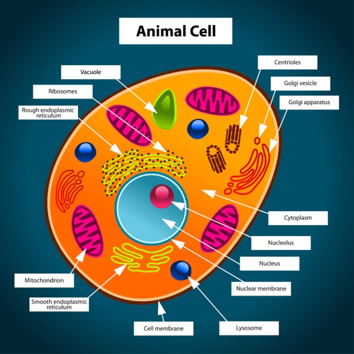
Animal Cell Structure
Animal cell coloring labeling worksheet – Animal cells are the fundamental building blocks of animal tissues and organs. Unlike plant cells, they lack a cell wall and chloroplasts, resulting in a more flexible structure and dependence on external sources for energy. Understanding their internal components is crucial to grasping the complexities of animal life.
Animal cells are eukaryotic cells, meaning they possess a membrane-bound nucleus containing the genetic material (DNA). Beyond the nucleus, a variety of specialized organelles perform distinct functions, working together to maintain cellular life and contribute to the overall organism’s health. The coordinated action of these organelles ensures the cell’s survival and proper functioning.
Key Organelles and Their Functions
The following table details the major organelles found within animal cells, their functions, and their typical shapes. Note that the precise shape of an organelle can vary depending on the cell type and its current activity.
Understanding animal cell structure is facilitated through activities like animal cell coloring labeling worksheets, which provide a hands-on approach to learning. These worksheets can be enhanced by incorporating visual aids that connect cellular components to real-world examples; for instance, exploring the diverse morphology of animals found in resources like free coloring pages of zoo animals can help students contextualize the functions of organelles within a broader biological framework.
Returning to the worksheets, the visual reinforcement aids in memorization and comprehension of complex cellular processes.
| Organelle | Function | Shape | Example/Detail |
|---|---|---|---|
| Nucleus | Contains the cell’s DNA; controls gene expression and cell activities. | Generally spherical, but can be irregular. | The nucleus acts as the cell’s control center, dictating protein synthesis and cell division. Its size varies depending on the cell’s activity. |
| Mitochondria | Generate energy (ATP) through cellular respiration. | Rod-shaped or oval, often described as bean-shaped. | Mitochondria are often referred to as the “powerhouses” of the cell. Their number can vary greatly depending on the cell’s energy demands. For example, muscle cells have many more mitochondria than skin cells. |
| Ribosomes | Synthesize proteins based on mRNA instructions. | Small, spherical structures. | Ribosomes can be found free-floating in the cytoplasm or bound to the endoplasmic reticulum. They are responsible for translating genetic code into functional proteins. |
| Endoplasmic Reticulum (ER) | Network of membranes involved in protein and lipid synthesis and transport. Rough ER (RER) has ribosomes attached; smooth ER (SER) lacks ribosomes. | Extensive network of interconnected tubules and sacs. | RER is involved in protein modification and transport, while SER synthesizes lipids and detoxifies certain substances. The extensive network facilitates efficient transport within the cell. |
| Golgi Apparatus (Golgi Body) | Processes, packages, and distributes proteins and lipids. | Stack of flattened, membrane-bound sacs (cisternae). | The Golgi apparatus acts as a sorting and packaging center, modifying and directing molecules to their final destinations within or outside the cell. |
| Lysosomes | Contain digestive enzymes to break down waste and cellular debris. | Small, membrane-bound sacs. | Lysosomes are crucial for maintaining cellular cleanliness and recycling cellular components. Their malfunction can lead to various cellular disorders. |
| Cytoskeleton | Provides structural support and facilitates cell movement. | Network of protein filaments (microtubules, microfilaments, intermediate filaments). | The cytoskeleton is a dynamic structure that constantly adapts to the cell’s needs, providing both stability and flexibility. |
| Cell Membrane | Encloses the cell; regulates the passage of substances into and out of the cell. | Thin, flexible membrane. | The cell membrane is selectively permeable, allowing only certain molecules to pass through. This is crucial for maintaining cellular homeostasis. |
Worksheet Design Considerations

Effective worksheet design is paramount for successful learning. A well-structured worksheet caters to diverse learning styles and skill levels, ensuring all students can engage meaningfully with the material. The design should facilitate understanding of animal cell structures and functions, not hinder it.A thoughtfully designed worksheet will improve comprehension and retention of information regarding animal cell components. Careful consideration of layout, visual aids, and labeling is crucial for achieving this goal.
Age and Skill Level Variations
Worksheet layouts should be adapted to suit the cognitive abilities and prior knowledge of different age groups. Elementary school worksheets should feature large, clear images, simple labeling, and minimal text. Activities could include coloring, matching, or simple fill-in-the-blank exercises. Middle school worksheets can incorporate more complex diagrams, require students to identify and label more structures, and include short-answer questions.
High school worksheets should present detailed diagrams, demand more in-depth labeling and analysis, and potentially include essay questions or research tasks. Furthermore, within each age group, variations in complexity should accommodate students with varying levels of understanding. For example, a simpler version might focus on identifying major organelles, while a more challenging version would include less common structures and their functions.
Effective Visual Aids
High-quality visuals are essential for conveying the intricate details of an animal cell. Microscopic images of real animal cells provide a realistic representation, allowing students to visualize the structures they are learning about. However, these images can be difficult to interpret for younger learners. Therefore, simplified, cartoon-style diagrams are more suitable for elementary school students. These diagrams should accurately represent the relative sizes and locations of organelles, while using bright, easily distinguishable colors to highlight individual components.
For older students, diagrams can be more detailed and include three-dimensional representations to enhance understanding of spatial relationships between organelles. Flowcharts illustrating cellular processes, such as protein synthesis or cellular respiration, can also be very effective visual aids. These visual aids should be meticulously labeled and referenced within the accompanying text.
Clear and Concise Labeling
Precise and unambiguous labeling is critical for successful learning. Labels should be clear, concise, and easy to read. Avoid using jargon or overly technical terms that students may not understand. For elementary students, labels should be simple and direct (e.g., “Cell Membrane”). For older students, labels can include more detail (e.g., “Phospholipid Bilayer of the Cell Membrane”).
The font size and style should be appropriate for the age group, with larger, bolder fonts used for younger learners. The use of consistent labeling conventions throughout the worksheet is also important to avoid confusion. For instance, always use the same color and font for the same organelle. Furthermore, a legend or key should be included to explain any abbreviations or symbols used in the diagram.
Creating the Coloring and Labeling Activity: Animal Cell Coloring Labeling Worksheet

This section details the creation of a visually engaging and educationally sound coloring and labeling activity for an animal cell diagram. The goal is to produce a worksheet that effectively reinforces student understanding of animal cell structure and function. A well-designed worksheet should be both informative and enjoyable for students to complete.The process involves designing a visually appealing diagram, selecting appropriate organelles for labeling, and organizing the labeling elements in a logical manner to facilitate student learning.
Careful consideration of the visual presentation and the sequence of labeling will significantly impact the worksheet’s effectiveness.
Animal Cell Diagram Design
The animal cell diagram should be a simplified, yet accurate, representation of a typical animal cell. The size and shape of the cell should be large enough to accommodate clear labeling of organelles without overcrowding. The overall shape should be roughly circular or oval, reflecting the typical morphology of an animal cell. Organelles should be depicted in their relative sizes and positions within the cell.
For example, the nucleus should be centrally located and relatively large compared to other organelles. The cytoplasm should be shown as a background filling the cell, surrounding all organelles. Mitochondria should be depicted as numerous, elongated structures scattered throughout the cytoplasm. The Golgi apparatus should be represented as a stack of flattened sacs, often near the nucleus.
The endoplasmic reticulum should be shown as a network of interconnected membranes, both rough (with ribosomes attached) and smooth. Lysosomes and ribosomes should be depicted as small, scattered dots within the cytoplasm. The cell membrane should form a clear boundary around the entire cell. The use of distinct colors for each organelle will enhance visual appeal and aid in identification during the labeling process.
Avoid excessive detail that might confuse students; the focus should be on the major organelles and their general arrangement.
Organelle Selection and Labeling Order, Animal cell coloring labeling worksheet
The worksheet should include the following organelles for labeling: nucleus, cytoplasm, cell membrane, mitochondria, ribosomes, endoplasmic reticulum (rough and smooth), Golgi apparatus, and lysosomes. Other organelles, such as centrioles or vacuoles (which are generally smaller in animal cells compared to plant cells), may be omitted to avoid overwhelming the students. The order of the labeling elements should be logical and progressive.
A suggested order would start with the largest and most prominent organelles, such as the nucleus and cell membrane, and then progress to smaller and more numerous organelles like mitochondria and ribosomes. This approach allows students to build their understanding of the cell’s structure in a stepwise manner. Providing a numbered list or a key with corresponding colors would further aid in organization and clarity.
